Neurofilament Light Chain (NfL)
A blood biomarker for neurodegeneration

NfL in the bloodstream
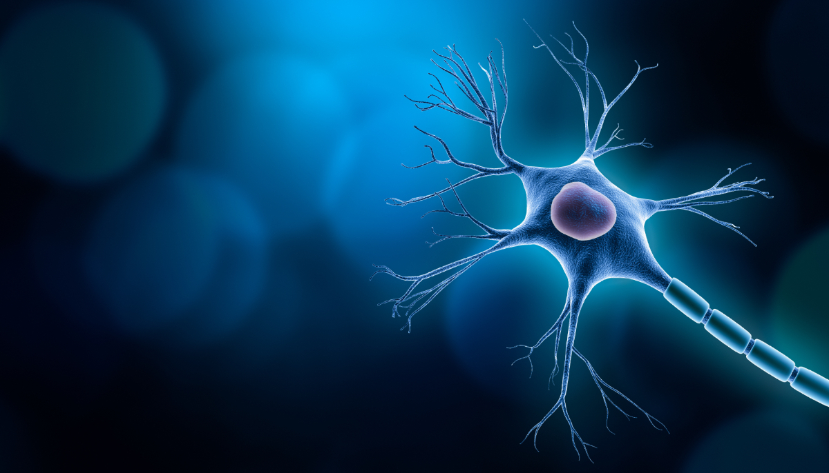
Neurofilament light chain (NfL) is a structural protein that normally helps support the shape and function of brain cells.1 In Alzheimer’s disease (AD), brain cells are damaged, which causes the brain to shrink (atrophy), and symptoms such as declining cognition to occur.2
When these brain cells are damaged, they release NfL into the bloodstream, which can then be detected in the blood plasma. So an increase of NfL levels in the plasma can indicate an increase in damage to brain cells.2
In fact, research has shown that blood plasma levels of NfL correlate directly with the severity of symptoms and pathology of AD.3,4 NfL levels have been shown to increase in preclinical AD, before symptoms appear, indicating neurodegeneration.5,6
An established biomarker
for brain damage
Increased amounts of NfL in the blood of those with AD corresponds to decreased thinking ability (cognition) and function.3,4 They also correlate with mild cognitive impairment (MCI) and other hallmarks of Alzheimer’s disease like brain shrinkage (atrophy) and tau tangles on a positron emission tomography (PET) scan.6
Moreover, high levels of NfL can predict how much a person with Alzheimer’s disease will decline in terms of their physical and mental capacity in the future.4 NfL is an established biomarker in Alzheimer’s disease, but it is not specific to Alzheimer’s disease.
Since NfL is released any time brain cells are damaged, that damage can be caused by other factors.6 Thus, NfL cannot be used for diagnosis of Alzheimer’s disease. However, in a person who has already been diagnosed, this protein may be used as a powerful tool for measuring and predicting disease progression.
-
References and useful links
1. Kadavath H, Hofele RV, Biernat J, et al. Tau stabilizes microtubules by binding at the interface between tubulin heterodimers. Proc Natl Acad Sci U S A. 2015;112(24):7501-7506. doi:10.1073/pnas.1504081112
2. Zetterberg H, Skillbäck T, Mattsson N, et al. Association of Cerebrospinal Fluid Neurofilament Light Concentration With Alzheimer Disease Progression. JAMA Neurol. 2016;73(1):60. doi:10.1001/jamaneurol.2015.3037
3. Ou YN, Hu H, Wang ZT, et al. Plasma neurofilament light as a longitudinal biomarker of neurodegeneration in Alzheimer’s disease. Brain Science Advances. 2019;5(2):94-105. doi:10.1177/2096595820902582
4. Raket LL, Kühnel L, Schmidt E, Blennow K, Zetterberg H, Mattsson-Carlgren N. Utility of plasma neurofilament light and total tau for clinical trials in Alzheimer’s disease. Alzheimer’s & Dementia: Diagnosis, Assessment & Disease Monitoring. 2020;12(1):e12099. doi:10.1002/dad2.12099
5. Preische O, Schultz SA, Apel A, et al. Serum neurofilament dynamics predicts neurodegeneration and clinical progression in presymptomatic Alzheimer’s disease. Nat Med. 2019;25(2):277-283. doi:10.1038/s41591-018-0304-3
6. Weston PSJ, Poole T, O’Connor A, et al. Longitudinal measurement of serum neurofilament light in presymptomatic familial Alzheimer’s disease. Alzheimer’s Research & Therapy. 2019;11(1):19. doi:10.1186/s13195-019-0472-5


